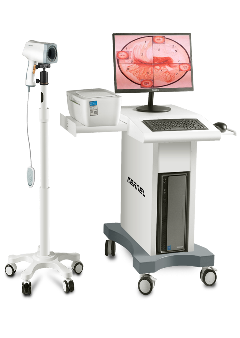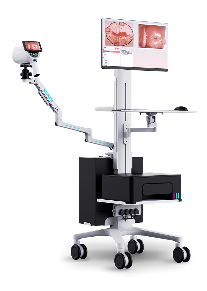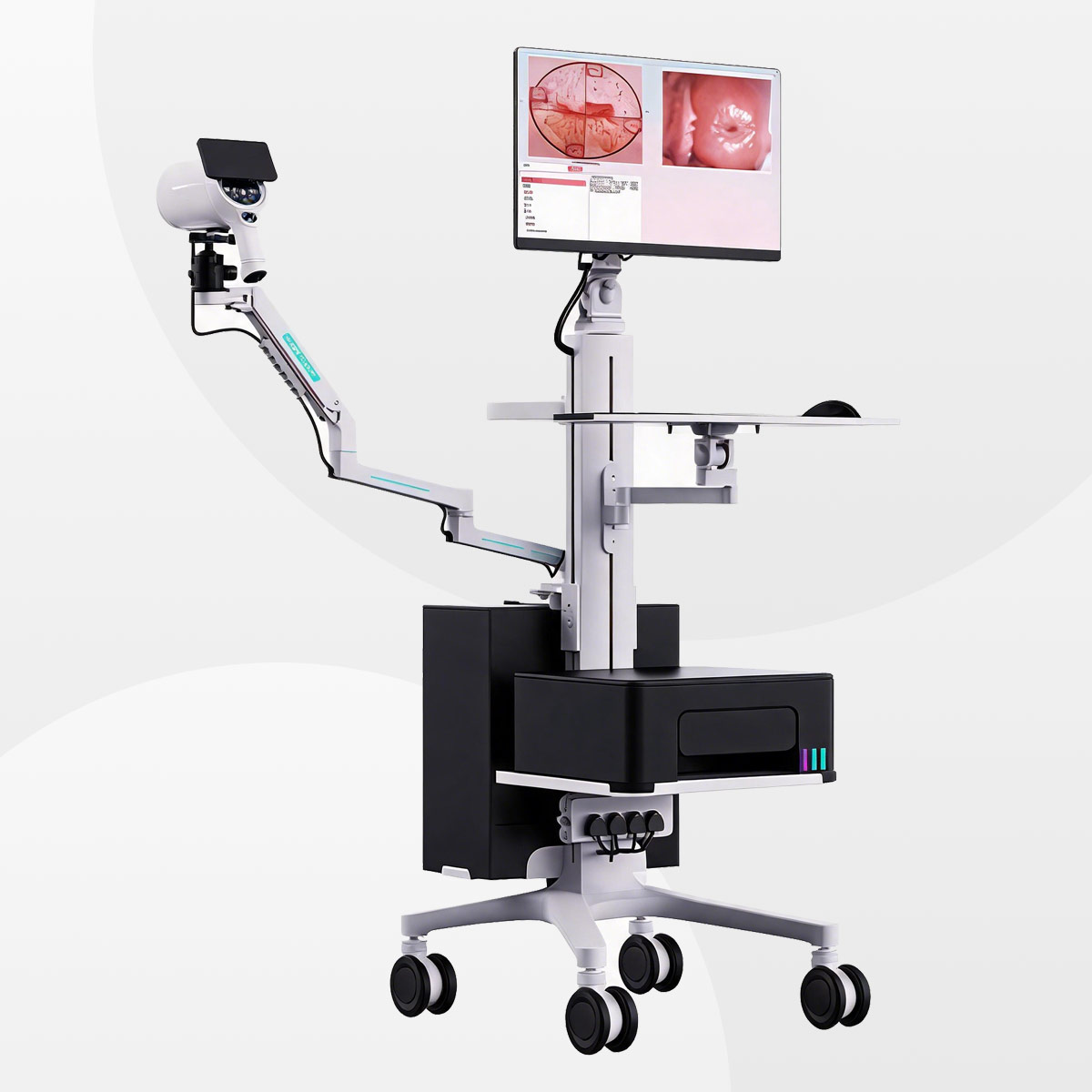
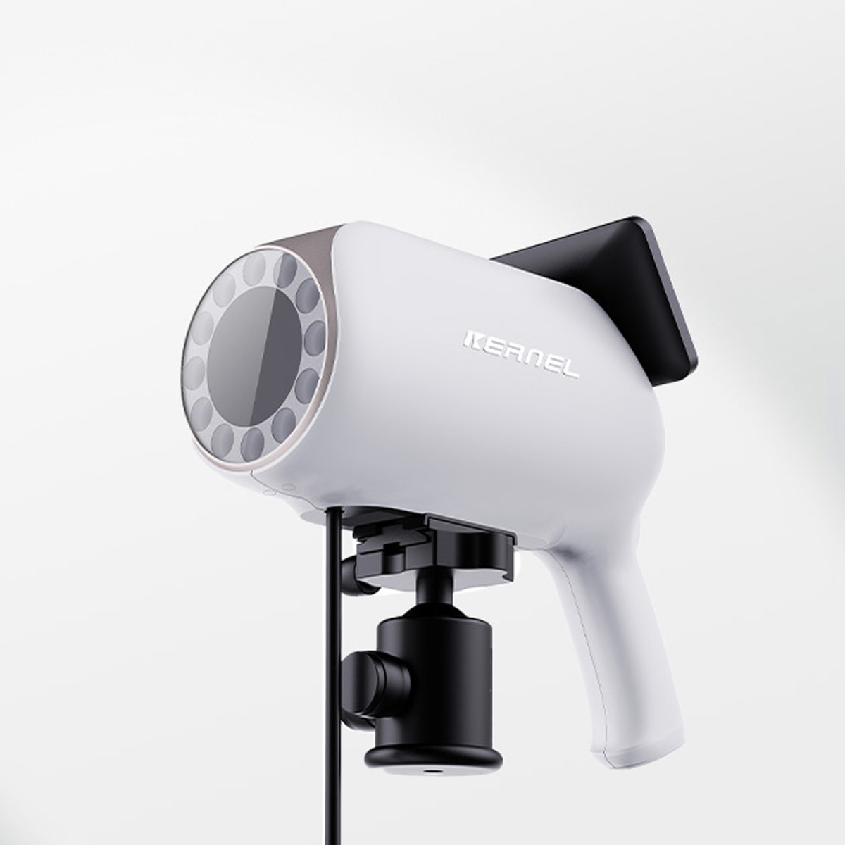
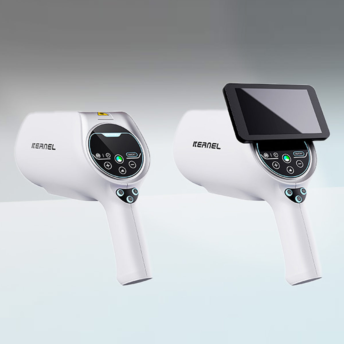
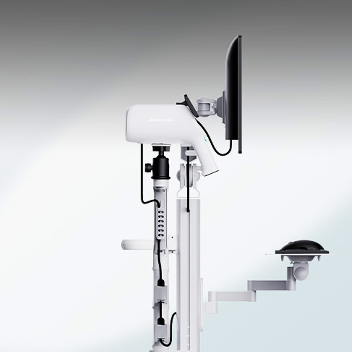
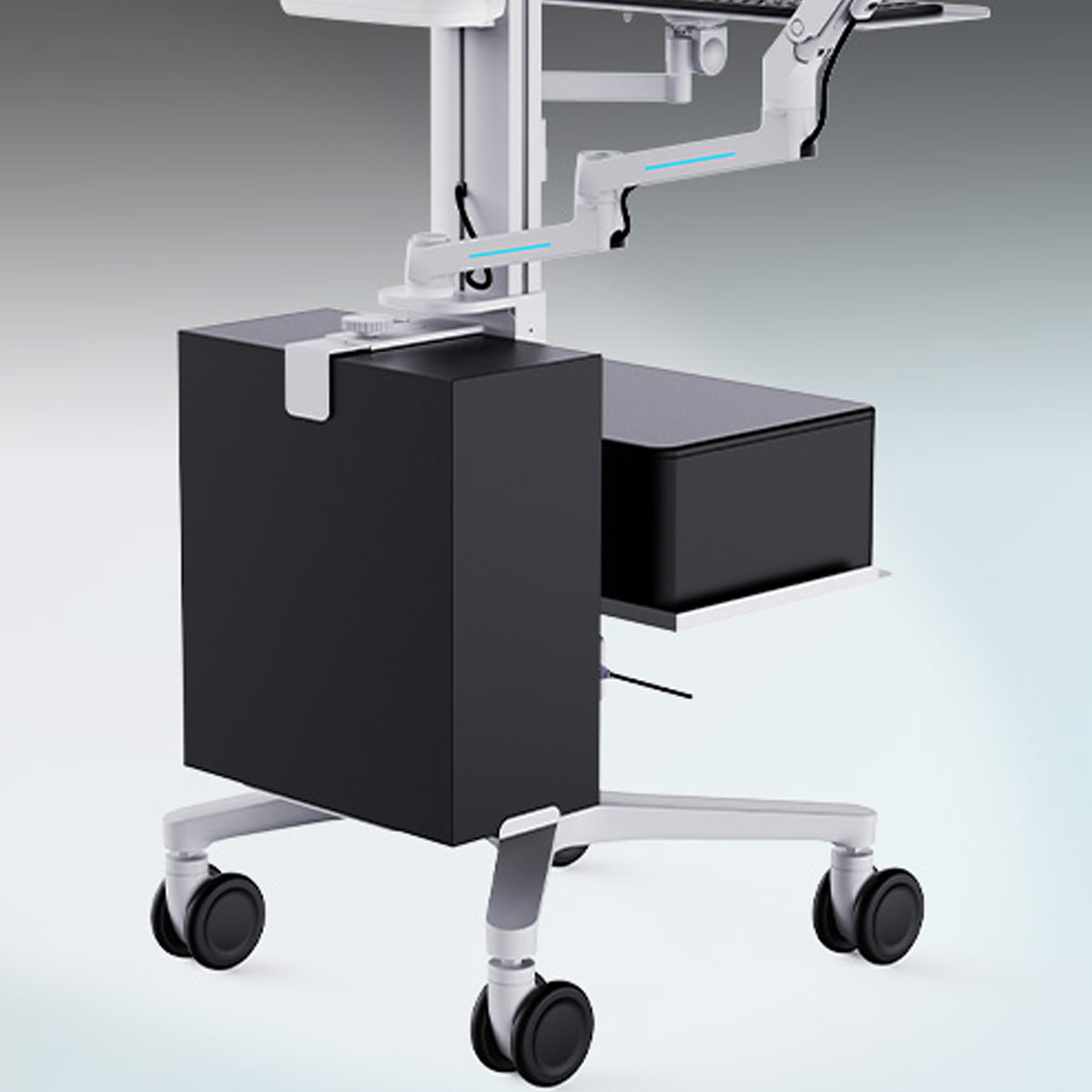
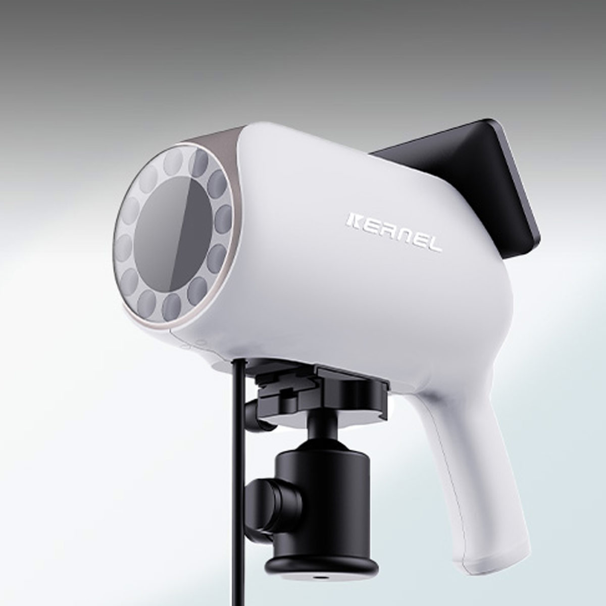
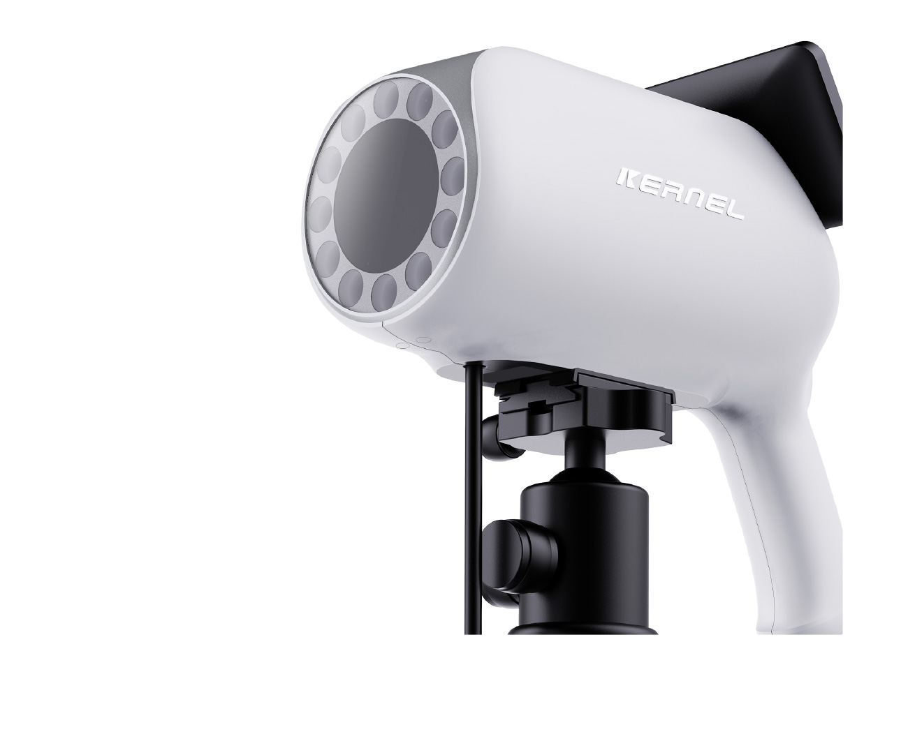
Digital HD Colposcope
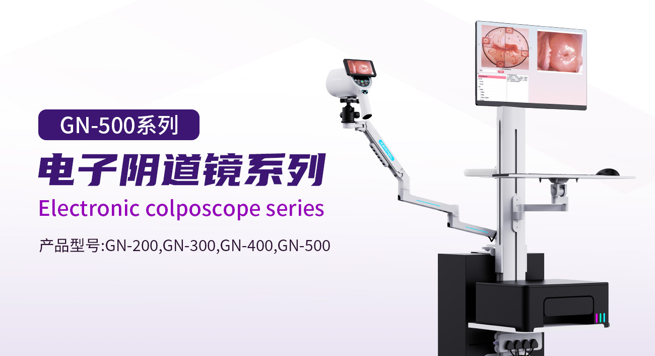
GN-500
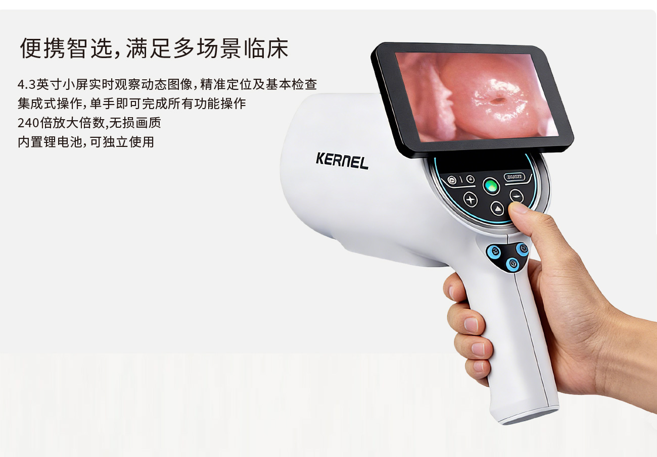
Used for clinical examination of the external genitalia, vagina, and cervix
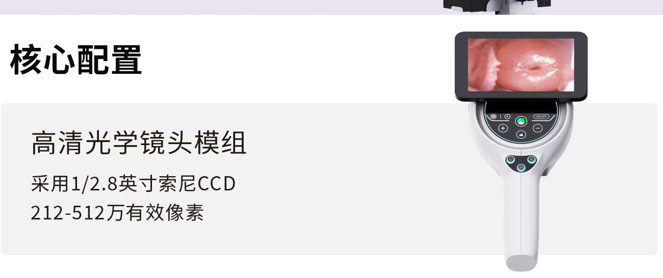
High resolution and high-definition digital CCD
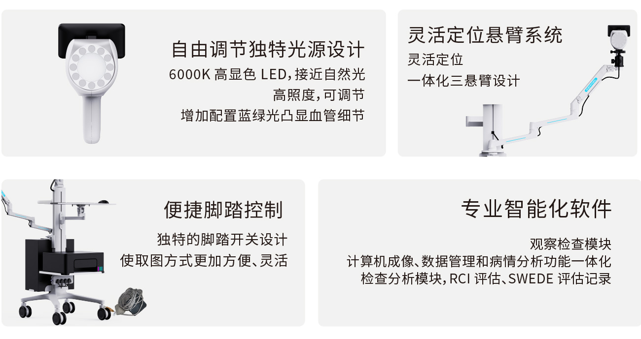
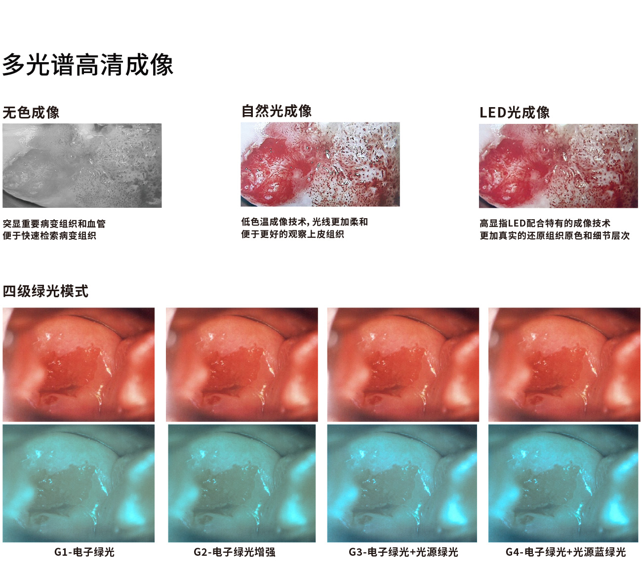
Free adjustment of unique light source design
The foot switch is flexible in taking pictures
Button and touch screen interaction
| Function Module | Specific Function Description |
|---|---|
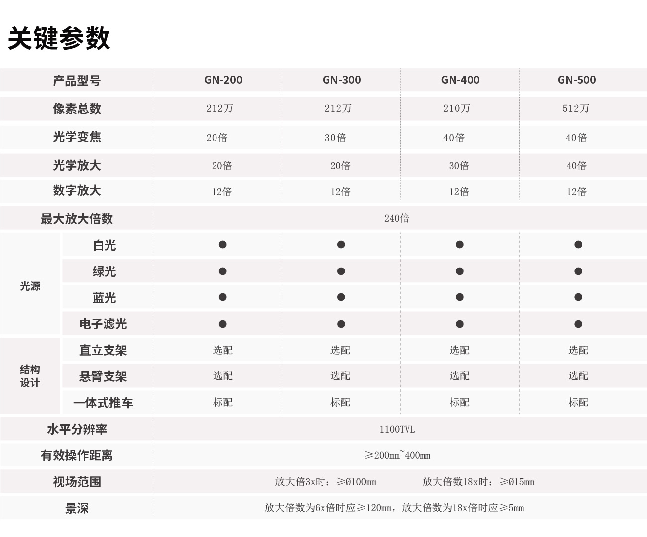
| Total Pixels | • ≥4 million |
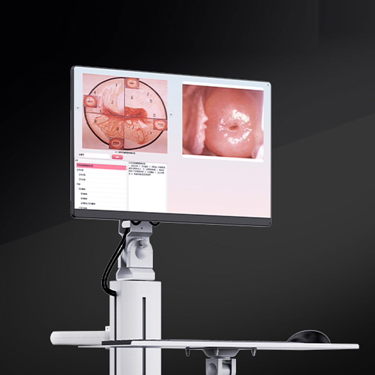
| Optical Magnification | • Has dynamic and static image comparison function, and can be compared with standard atlas • Captured images can be enlarged, reduced, converted to negative, marked, text-annotated, locally magnified, and used for area and perimeter calculation, etc., facilitating doctors in identifying lesion tissue |
| Horizontal Resolution | • ≥1100TVL |
Q1: What main models are available in the Komed KN‑2200 series, and how are they differentiated?
A: They are divided into two major series: electronic digital models and optical-electronic integrated models:
1. KN‑2200 / KN‑2200A / KN‑2200Ⅰ / KN‑2200Ⅰ(H) / KN‑2200Ⅰ(4K): pure electronic digital colposcopes; KN‑2200Ⅰ(4K) is the 4K ultra-HD version with higher image resolution, suitable for cervical specialty screening in tertiary hospitals and maternal and child health hospitals.
2. KN‑2200B / BⅠ / BⅡ: optical-electronic integrated colposcopes with a binocular optical microscope + electronic imaging dual channel; they allow both eyepiece optical observation and monitor image review, with strong stereoscopic perception, suitable for pathological biopsy and surgical operation scenarios at Komed Medical.
Q2: What are the core differences between the optical-electronic integrated B series and ordinary electronic colposcopes?
A: The B series is equipped with a binocular stereoscopic optical microscope and has a direct optical observation channel, with optical magnification, large depth of field, and strong stereoscopic perception of lesions, facilitating intraoperative targeted biopsy; electronic models observe only through a monitor, have a simple configuration, and are suitable for large-volume preliminary screening in outpatient clinics at Komed Medical.
Q3: What core components does the system include?
A: Camera host / optical microscope, damped cantilever stand, foot pedal capture switch, control handle, software system; computer, monitor, and printer are optional peripherals.
Q4: What clinical functions does the software have?
A: Image freeze capture, dual capture via foot pedal and handle, lesion annotation, image comparison and analysis, multiple sets of gynecological report templates, medical record storage/query/statistics, report printing output.
Q5: What clinical scenarios is the colposcope suitable for?
A: It is used for magnified examination of the vulva, vagina, and cervix; suitable for abnormal TCT/HPV screening, visually suspicious cervical lesions, postcoital bleeding, follow-up after cervical treatment, preoperative assessment of lesion extent, assisting cervical lesion screening, and providing a basis for biopsy localization.
Q6: Can a colposcope directly diagnose cervical cancer?
A: No. A colposcope is a magnified imaging examination device used to identify suspicious lesions and guide precise biopsy; the final diagnosis depends on pathological biopsy results.
Q7: What is the examination procedure?
A: The patient assumes the gynecological position, a speculum is inserted, the device is adjusted to a suitable working distance of 200‑400 mm, and light source brightness is adjusted; observations are made sequentially with saline, acetic acid, and iodine tests; images are captured via foot pedal / handle; suspicious points are marked, and biopsy is performed if necessary; report saving and printing are completed.
Q8: Is the device compatible with biopsy surgical operations?
A: The KN‑2200B optical-electronic integrated model has binocular optical observation, and the cantilever can hover at multiple angles, making it suitable for outpatient biopsy and minor surgical operations; electronic models are mostly used for imaging screening.
Q9: What are the requirements for working distance and light source illuminance?
A: Standard working distance is 200‑400 mm; light source illuminance ≥1200 Lux to ensure sufficient image brightness for long-distance observation.
Q10: What are the features of the cantilever stand?
A: Fully damped pan-tilt cantilever, 360° multi-angle free hovering, flexible adjustment of camera position and angle, freeing both hands, and adapting to different examination positions.
Q12: What is the function of the foot switch?
A: It allows rapid capture and saving of images by foot during examination without touching the host mouse, making doctor operations smoother.
Q13: Is the device an invasive instrument? What parts need disinfection?
A: The device host and camera lens do not enter the human body; the lens should avoid contact with human tissue; the speculum is an independent consumable; the lens housing should be wiped with disinfectant wipes; liquid should not be poured directly onto the device.
Q14: What are the daily maintenance points for the device?
A: Avoid bumping the lens; wipe the lens housing with a dust-free soft cloth; do not directly wipe the camera lens with alcohol; avoid humid environments during use; regularly check stand damping and light source brightness; regularly back up the medical record database in the software.
Q15: What are the warranty and after-sales policies?
A: The standard warranty for the whole machine is 1 year; within the warranty period, non-human-caused quality problems are repaired or replaced free of charge; the manufacturer provides installation training and clinical operation training; after the warranty period, long-term technical support is provided, and extended warranty service is available. Shanghai Jumu Medical...
Q16: Can it connect to hospital HIS/LIS systems?
A: The software supports interface integration and can be customized according to hospital needs to achieve medical record data interoperability.